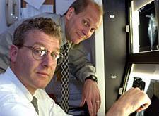
Lead author, Prof. Steven
Poplack & co-author,
Keith D. Paulsen
(click to enlarge)
A new study in Radiology (journal of the Radiological Society of North America) published by Dartmouth physicians and engineers uses results from a five-year project that tested three new imaging techniques to examine breast abnormalities that include cancer.
The study found that new methods of electromagnetic imaging offer a high contrast and are able to distinguish between healthy and abnormal breast tissue.
Ninety-seven of the 150 women who participated in the study had an abnormal conventional breast image that was found to be suspicious or highly suggestive of malignancy. Prior to biopsy, these women underwent electromagnetic exams that allowed researchers to compare the abnormal area with the background breast tissue and with a mirror image area in the opposite breast and correlate the data with biopsy findings. Upon further analysis, the researchers were able to determine that the new imaging techniques provided an increase in contrast between 150 and 200 percent to discriminate between breast cancer and benign tissue.


Ad Statistics
Times Displayed: 45316
Times Visited: 1411 Keep biomedical devices ready to go, so care teams can be ready to care for patients. GE HealthCare’s ReadySee™ helps overcome frustrations due to lack of network and device visibility, manual troubleshooting, and downtime.
According to lead author Steven Poplack, associate professor of radiology and OB/GYN at Dartmouth Medical School and co-director for breast imaging/mammography at DHMC, the new imaging techniques were put to the test to quantify their effectiveness, and the results showed the potential power of using a variety of imaging techniques to get the best possible view of what's going on in the breast tissue.
The electromagnetic techniques are electrical impedance spectral imaging (EIS), microwave-imaging spectroscopy (MIS) and near infrared (NIR) spectral imaging. The three techniques demonstrated significant differences in region-of-interest image summaries of normal versus abnormal breasts for EIS, across diagnostic groups for NIR, and for MIS when analysis was restricted to lesions larger than one centimeter. When all were used together, the electromagnetic imaging modalities seemed to be even more accurate.
EIS is a painless test that uses a low voltage electrode system to examine how the breast tissue conducts and stores electricity. Different cancer cells have different electrical characteristics, and living cell membranes carry an electric potential that affect the way a current flows.
The MIS exam is a technique that is particularly sensitive to water and involves the proliferation of very low levels (1,000 times less than a cell phone) of microwave energy through breast tissue to measure electrical properties. Tumors have been found to have more water and blood than regular tissue.


