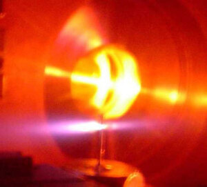
Duke's new technology
uses two lasers pulsing
a few femtoseconds
at a time
(click to enlarge)
The distributions of hemoglobin, a component of red blood cells, and melanin, a skin pigment, serve as early warning signs for skin cancer growth. But because skin scatters light strongly, simple microscopes cannot be used to locate those molecules except right at the surface. Although laser methods have been developed to probe deeper down for some other molecules that can be made to glow, both melanin and hemoglobin remain dark and inaccessible using those methods.
"The standard way physicians do a diagnosis now is to cut out a mole and look at a slice of it with a microscope," said Warren Warren, the James B. Duke Professor of Chemistry, Radiology and Biomedical Engineering, and Director of Duke's new Center for Molecular and Biomedical Imaging. "What we're trying to do is find cancer signals they can get to without having to cut out the mole. This is the first approach that can target molecules like hemoglobin and melanin and get microscopic resolution images the equivalent of what a doctor would see if he or she were able to slice down to that particular point," Warren said.
Warren's group has now developed a technology for coaxing both hemoglobin and melanin inside questionable skin moles to emit light by exciting them with highly controlled laser pulses. The innovation uses a delicate interplay between two laser beams, each emitting a different color of light. To keep the skin from overheating in the process, the lasers must also be able to pulse on for only femtoseconds -- a thousand trillionths of a second -- at a time.


Ad Statistics
Times Displayed: 2045
Times Visited: 23 Keep biomedical devices ready to go, so care teams can be ready to care for patients. GE HealthCare’s ReadySee™ helps overcome frustrations due to lack of network and device visibility, manual troubleshooting, and downtime.


