by
John R. Fischer, Senior Reporter | April 11, 2022
From the April 2022 issue of HealthCare Business News magazine
While a growing field, Dr. Martin Goldman, Dr. Arthur Master Professor of Medicine/cardiology and vice chair of cardiology at Mount Sinai Heart in New York, says the value of the information is directly related to quality of images, which is directly correlated to level of training for personnel performing the imaging. “The ability to share the images via telehealth for doctor consultations is very good. But the key thing with the echo is garbage in garbage out. You can really distort the image if your acquisition is very poor, and as a result, distort the interpretation.”
Expanding reach
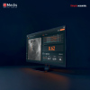
Historically used in cath labs and medical imaging suites, cardiac ultrasound can be found today in a range of places, from emergency rooms to intensive care units to even primary care physicians offices. This is thanks in part to the rise of point-of-care ultrasound (POCUS), which has also made it accessible in ambulances, paramedic units, helicopter medical flight programs and even on battlefields by non-cardiology personnel.
Ranging from carts to handheld devices, the technology can scan patients right at their bedside for faster exams and assessments of individuals who are challenging to move to imaging suites. “Because you can do it bedside, you don’t have to move the patient to another room. You can quickly move from patient to patient and disinfect in between patients. It really assists in how we diagnose and assess patients to improve care,” said Krys Lee, clinical analyst for symplr and registered ultrasound technologist.
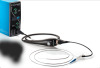
In critical care units and emergency rooms, providers are also starting to perform transesophageal echo (TEE) where a miniature echo probe is attached to a long thin tubing unit that is inserted down the patient’s throat to visualize heart function, valves and potential effusion. This provides a closer and precise view compared to transthoracic echo (TTE), where the probe is placed on a person’s chest. “They actually make small, thin, long probes now, that can be put into the esophagus during chest compressions if the patient has a cardiac arrest, to help better understand the etiology of the low blood pressure of EKG abnormalities and expedite appropriate therapeutic intervention,” said Goldman.
Another new use is Focused Assessment with Sonography in Trauma (FAST) exams to look for blood around the heart, assess the heart and look for blood around other organs at the bedside. These allow for cardiac ultrasound to assess a wider range of conditions and spares patients more often from costly imaging.