by
Lauren Dubinsky, Senior Reporter | June 05, 2017

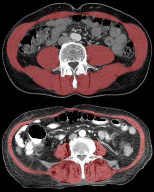
CT image showing reduced size
and density of core muscle
(highlighted in red)
CT imaging has been used to predict patient outcomes after a hip fracture, but radiologists at UC Davis and Wake Forest Baptist medical centers were the first to use the technology to guide treatment decisions.
"As patients age, it becomes increasingly important to identify the safest and most beneficial orthopaedic treatments, but there currently is no objective way to do this," Dr. Robert Boutin, professor of radiology at UC Davis, said in a statement. "Using CT scans to evaluate muscles in addition to hip bones can help predict longevity and personalize treatment to a patient's needs.”
If the CT exam reveals that the patient has a favorable life expectancy, they could be treated with total hip arthroplasty, which could lessen the chance of re-operation, and improve hip function and quality of life. Alternatively, if the exam shows evidence of frailty, they could benefit most from a simpler surgery.


Ad Statistics
Times Displayed: 16169
Times Visited: 33 Final days to save an extra 10% on Imaging, Ultrasound, and Biomed parts web prices.* Unlimited use now through September 30 with code AANIV10 (*certain restrictions apply)
The study involved almost 3000 patients who were at least 65 years old and treated for fall-related injuries at UC Davis between 2005 and 2015. It was suspected that all of them had broken hips, so CT exams were ordered to diagnose or rule out fracture.
Boutin and his team evaluated the CTs as well as additional measurements of the size and density of lumbar and thoracic muscle alongside the spine. The information was then compared with mortality data from the National Death Index, which is the CDC’s centralized database of death record information.
They found that patients with CT scans that showed stronger core muscle had substantially better survival rates over the course of the 10-year study.
Senior author of the study, Leon Lenchik, described these findings as “remarkable.” The majority of the previous research on CTs of muscle has been focused on cancer patients and involved larger sample sizes.
Boutin and Lenchik hope this research will encourage other research teams to study muscle loss, which is a global epidemic. They also hope this will lead to better orthopedic treatments for the growing elderly population.

