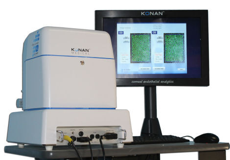
The CellChek XL™ delivers cellular level imaging of the corneal endothelium with the industry’s most comprehensive tools for cellular morphology and trending analysis. The system is used globally for routine clinical use including many of the leading medical and surgical applications such as: ICLs (FDA labeling requires), cataract surgery and premium IOLs, glaucoma medication assessment, DSEK / DMEK, keratoconus, and corneal cross-linking (CXL). Many clinicians also use specular microscopy for clinical assessment of contact lens related corneal distress.
As a fully automated system, the CellChek XL includes non-contact pachymetry and peripheral analysis features critical for following tissue transplantation procedures. The microscope system includes 19″ touch screen, motorized table, and printer. Konan provides on-site training, advanced topics web-enabled training and the industry’s best clinical support.
the industry’s gold standard “Center Method™”
location data captured, critical for trends analysis
one of the highest diagnostic procedure reimbursements
high ROI
easy to use, easy to interpret
technician administered, 1 minute test
own the market leader and gold standard
Return Policy
Items are sold as-is with no returns or refunds available unless explicitly stated.

 Get a CPS 2-Year Protection Plan starting at $5.99. Sold separately. Learn more