New MRI Developed by Research Team
by
Joan Trombetti, Writer | June 25, 2008

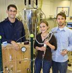
Leif Schroeder (left)
and team members
Researchers at the Department of Energy's Lawrence Berkeley National Laboratory, and the University of California at Berkeley, have developed a magnetic resonance imaging method that is faster, more selective, and many thousands of times more sensitive than standard MRI.
Using "temperature-controlled molecular depolarization gates," the new technique builds on a series of previous developments in MRI, and the closely related field of nuclear magnetic resonance (NMR).
Standard MRI suffers from low sensitivity and requires patients to remain motionless for long periods of time inside noisy, claustrophobic machines. The new method requires less time and is able to distinguish even among specific target molecules.
"The new method holds the promise of combining a set of proven NMR tools for the first time into a practical, supersensitive diagnostic system for imaging the distribution of specific molecules on such targets as tumors in human subjects or even on individual cancer cells," said lead author Leif Schroeder of Berkeley Lab's Materials Sciences Division.
MRI and NMR make use of the quantum-mechanical phenomenon known as nuclear spin; nuclei with odd numbers of protons or neutrons will orient themselves like tiny bar magnets, spin "up" or spin "down," in a strong magnetic field. If the spinning nuclei are knocked off-axis by a jolt of radio-frequency (RF) energy, they wobble or precess at a characteristic rate, a rate that is strongly conditioned by their immediate chemical neighbors. During a certain relaxation time (typical of each atomic species in a specific environment), the nuclei reorient themselves and emit a radio signal that reveals both their position and their chemical surroundings.
The spin-up state requires fractionally less energy, so there's typically a slight excess of spin-up nuclei, about one in a hundred thousand (.001 percent) and it's this tiny difference that yields a useful signal. In clinical settings MRI is usually done using hydrogen nuclei, protons, which are ubiquitous in the human body. But other nuclear species, notably the noble gas xenon, offer advantages over hydrogen that in the case of xenon include a virtual absence of background signal, since there is no xenon in biological systems.
Xenon is useful in MRI and NMR because the spins of its nuclei are readily polarized, in a process involving contact with rubidium vapor irradiated with a laser beam. In such "hyperpolarized" xenon, the excess of spin-up nuclei can be as much as 20 percent, which gives a far stronger signal than hydrogen's .001 percent spin-up excess. Hyperpolarized xenon has a much longer relaxation time than hydrogen.
