Breast MRI Holds Promise
by
Joan Trombetti, Writer | March 31, 2008
MRI detected more
than double the number
of cases of DCIS
than mammography.
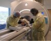
Since early detection is a key in women diagnosed with breast cancer, it has become obvious that MRI (Magnetic Resonance Imaging) can play a significant role in breast imaging and is gaining momentum among researchers because it has been found to be superior to mammography whether for diagnosing primary or recurrent, invasive or intraductal, familial or sporadic breast cancer, regardless of the density of a woman's breast.
MRI can take pictures of both soft and hard breast tissue, generating cross-sectional images in all planes, including oblique and can distinguish diseased tissues like tumors and lesions. It has mostly been used for brain imaging and has long been considered less sensitive than mammography for detecting ductal carcinoma in situ (DCIS), a precursor to breast cancer that is confined to the milk ducts.
A study presented by Professor Christiane K. Kuhl at the European Congress of Radiology (ECR) recently showed that MRI detected more than double the number of cases of DCIS than mammography, especially high-grade DCIS the most aggressive form of the condition. Out of 167 women imaged and diagnosed with pure DCIS, 56 percent were diagnosed by mammography and 92 percent by MRI, the study revealed. Of the 89 high grade DCIS, 43 (48 percent) were missed by mammography but diagnosed by MRI alone. By contrast, MRI detected 87 (98 percent) of these lesions and the two cases missed by MRI were detected by mammography.
The American Cancer Society (ACS), reports that breast cancer mortality has been dropping significantly since 1990 because of better treatments and earlier detection, and mammography dramatically improves detection at an early stage. But, Professor Kuhl says that breast cancer is still one of the most frequent cancers overall and continues to be one of the leading causes of death in women, indicating that there is room and need for improvement. Kuhl explained that breast MRI has had a reputation of causing too many false positive results that have incorrectly suggested the presence of cancer. However, she believes that this is due to the level of expertise with which the average breast MRI study had been read - compared to the average level of expertise with which a mammogram is interpreted. She points out that more recent results of large multicenter trials on breast MRI suggest that the PPV (true-positive biopsy rate) of breast MRI is indeed similar to or even higher than that achieved with mammography.
Lack of technical standards, trained radiologists, quality assurance programs and minimally invasive biopsy capabilities discourage clinicians from using breast MRI, and Kuhl believes that all of these difficulties can be easily solved if more breast MRI studies were done.
Standards are currently being developed and MR-guided, vacuum-assisted biopsy for minimally invasive tissue diagnosis is already available. But this system is expensive, such that the investment will only pay off for institutions that see enough patients for breast MRI, according to Kuhl.
Kuhl also feels that the alleged lack of evidence by randomized controlled clinical trials and fear of over-treatment are used to discourage the use of breast MRI for staging women diagnosed with breast cancer and the time has come to discuss these concerns and weigh them against the advantages of screening and diagnostic applications of breast MRI.