by
Brendon Nafziger, DOTmed News Associate Editor | May 31, 2011

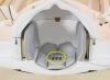
A 20-year-old's
pelvis, in pink, is
almost one inch thinner
than a 79-year-old's,
in black. (Image credit:
Laurence Dahners, M.D.)
The expanding girth most people associate with fat building up in middle age might be caused partly by bone growth, according to researchers.
Although most adults stop growing in height by the time they hit 20, researchers examining CT scans from hundreds of patients have discovered their hipbones keep growing as they age.
In fact, in the study published online last week in the Journal of Orthopaedic Research, researchers found patients in their 70s had pelvises on average one inch larger than patients in their 20s. This could translate into a three-inch waist size increase from age 20 to 79, the researchers said.


Ad Statistics
Times Displayed: 186622
Times Visited: 4999 For those who need to move fast and expand clinical capabilities -- and would love new equipment -- the uCT 550 Advance offers a new fully configured 80-slice CT in up to 2 weeks with routine maintenance and parts and Software Upgrades for Life™ included.
And if the whole body is widening at the same rate, it could explain some of the one pound a year weight gain people experience during this period.
"Our findings suggest that pelvic growth may contribute to people becoming wider and having a larger waist size as they get older, whether or not they also have an increase in body fat," Dr. Laurence E. Dahners, senior author of the study and a professor of orthopaedics with the University of North Carolina School of Medicine in Chapel Hill, said in a statement.
For the study, the researchers examined CT scans of 246 randomly selected patients between 20 and 79, with 20 male and 20 female patients in each 10-year age group. The researchers used the scans to gauge the width and height of the L4 vertebral body, the width of the pelvic inlet and the distance between the hip joints and their diameter.
According to a UNC summary of the study, the scans showed the width but not the height of the L4 vertebral body, the width of the pelvic inlet and the distance between the hip joints, and the diameter of the hip joints all increased with age.
Dahners said the pelvic inlet widening was also evidence of true pelvic growth, as the inlet would be smaller if appositional bone formation occurred.