by
Brendon Nafziger, DOTmed News Associate Editor | February 08, 2011
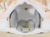
Researchers admire their
results, with the
MRI-HIFU scanner in
the background. (Photo courtesy
Philips Research)
A system combining MRI and ultrasound technology might be able to better direct chemotherapy drugs to tumors while sparing healthy tissue, in a breakthrough that could one day improve cancer treatments.
On Monday, researchers affiliated with Royal Philips Electronics and Eindhoven University of Technology in the Netherlands said they had completed proof-of-concept work in rats showing how the two modalities could work together to more precisely deliver drugs while also measuring the exact amount of drug absorbed by the tumor.
In the experiment, ultrasound is used to "activate" cancer-killing drugs at the site of tumors, thereby delivering a bigger cancer-fighting payload where it's most useful, Philips said. And the MRI imaging is used to spot the tumor and track how much of the cancer-killing compounds have been taken up, to check if the therapy works.


Ad Statistics
Times Displayed: 186660
Times Visited: 5008 For those who need to move fast and expand clinical capabilities -- and would love new equipment -- the uCT 550 Advance offers a new fully configured 80-slice CT in up to 2 weeks with routine maintenance and parts and Software Upgrades for Life™ included.
The research, led by Holger Grüll, a professor at Eindhoven who also does molecular imaging research for Philips, is to be published this month in the Journal of Controlled Release.
Just right
Getting the right dosage for chemotherapy is tricky: doctors want to use a high dose to kill tumors. But since the drugs attack most rapidly dividing cells, and not just cancerous ones, they can have serious side effects, such as anemia and increased risk of infection. Because of this, doctors can't use as high a dose as might be effective.
But the project, part of the European Union-funded "sonodrugs" program which looks at ways to better deliver medicine, hopes to improve on this.
The gist is to combine chemotherapy drugs with fat particles called liposomes, which become leaky at high temperatures. For the study, the drug doxorubicin was encapsulated in the liposomes, Philips said in materials posted on its website.
At the normal body temperature of 37 degrees Celsius, the liposome stays intact, sealing in its drug payload and protecting healthy tissue, Philips said.
[
Read about Philips and Celsion's earlier work on ultrasound-activated cancer drugs. ]
And this is where ultrasound comes in: the researchers use a type of heat-generating ultrasound, called a high intensity focused ultrasound (HIFU) beam, which heats up the tumor region to 42 degrees Celsius. This causes the liposomes to leak, letting their drug contents escape.
Measure for measure
To pinpoint the tumor to guide the HIFU beam, the doctors use MRI imaging. This modality also helps capture the tumor's temperature during the heating process to ensure it's evenly heated to 42 degrees Celsius (plus or minus about 1 degree, according to Philips).