Philips scanner will
digitize slides in under a minute.
Philips Healthcare is teaming up with Danish software company Dako to provide analysis programs for its forthcoming pathology slide scanner, the med tech giant said Wednesday.
The announcement comes only several months after Philips struck a deal with Definiens, a Munich, Germany-based imaging analysis company, to start a project investigating support tools for breast cancer diagnosis with the device.
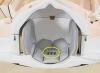
The scanner, due within the next six months, can digitize a pathology slide in less than a minute, making it one of the fastest such scanners available with the potential to expand what until now has been a limited market, Philips said.


Ad Statistics
Times Displayed: 370330
Times Visited: 8746 Quality remanufactured Certified Centrifuges at Great prices! Fully warranted and backed by a company you can trust! Call or click for a free quote today! www.Centrifugestore.com 800-457-7576
Generally, pathology labs chemically "fix" samples on glass slides, and then stain them with biological products or chemicals to make it easier to visualize certain structures or proteins in order, for instance, to help with a cancer diagnosis once the slide is put under a microscope.
By digitizing samples, labs could have an easier time tracking them down in an archive, Philips said. Typically, a big lab will process hundreds of samples a day.
"Retrieving a particular sample among a jungle of samples, you can imagine if you were to do it digitally, it could be done more efficiently," Steve Klink, a spokesman for Philips Digital Pathology, told DOTmed News.
Scanning and digitizing pathology slides could also make it easier to share findings with pathologists in remote locations. And this is where Dako comes in; pathologists could use software to quantify image analysis and help diagnose diseases, much as some radiologists use automated programs to help find tumors on a mammogram.
Philips hopes the scanner, which debuted at the United States and Canadian Academy of Pathology's annual meeting earlier this year, will fit easily into a pathologist's workflow, as it can capture an image in less than one minute. One of the obstacles to adoption of the technology in the past has been the slow speed of scanners, Philips said, making them impractical to use in high-volume labs.
Pathology scanning's slowness comes, in part, from the massive size of pathology slide files. A 1.5 by 1.5 centimeter slide, when digitized, can become a nearly 10-gigabyte file, Philips said, exceeding even the size of the memory-hogging file of a CT scan.
"It's because of the amount of details," Klink said. He compares the amount of detail that needs to be preserved in a digital pathology slide to a photograph of a stadium from a helicopter where you could zoom in to see each individual blade of grass with near perfect clarity.
Dr. Clive Taylor, a pathologist and professor at University of Southern California in Los Angeles, welcomed the announcement.
"It is exciting that collaborations like that between Dako and Philips are now bringing diverse but appropriate expertise to bear on implementing a full digital pathology program," he said in a statement provided by Philips.
The scanner with the image management system should launch within the next six months, Philips said, while integration with the Dako imaging tools should occur by the second half of 2011.
Initially, the as yet unnamed scanner will only be sold for research, as it will need U.S. Food and Drug Administration approval before it can be used for cancer diagnoses. Philips said it's looking to focus initially on Dako's breast cancer diagnosis tools, and then move onto prostate and colon cancer.