Thanks to Cancer's Inflexibility, an Ultrasound Tool Can Better Detect Breast Lesions
by
Brendon Nafziger, DOTmed News Associate Editor | December 04, 2009

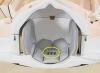
Elasticity software was
applied to this ultrasound
image and the solid mass
was noticeably larger
than without elasticity software
An ultrasound technique that measures stiffness of tissue dramatically improves detection rates in finding breast cancer, according to a presentation given Monday at the annual meeting of the Radiological Society of North America (RSNA).
Researchers at the Elizabeth Wende Breast Care center in Rochester, New York found that elastography, which uses ultrasound to determine how elastic tissue is, was able to heavily reduce the number of false positives in breast cancer cases, which often lead to unnecessary biopsies. In fact, recent studies estimate that only one out of 10 biopsies performed following ultrasound finds cancers.
In the current study, the doctors looked at 198 suspicious findings from 193 patients, of which around 140 were biopsied. Of the 140 biopsies, they discovered 59 cancers.
As expected, elastography was incredibly sensitive, according to the researchers. "Elastography correlated with needle biopsy results 98 percent of the time, for 58 out of the 59," Stamatia Destounis, M.D., the lead author of the study and a doctor at the clinic, tells DOTmed News.
But, equally important, it appeared to be fairly specific. "Of the 69 benign findings, the correlation with elastography was 78 percent." That is, it misidentified harmless growths as cancerous, or was simply unclear, in only 22 percent of the cases, an error rate far lower than that found in traditional -- and fairly unspecific -- ultrasound.
Cancers are inelastic
Elastography works because scientists believe that cancerous tissue is firmer than the surrounding healthy breast tissue. When images from elastography are viewed side by side with conventional ultrasound images, malignant lesions appear larger.
"This is something people have worked on for a while," Dr. Destounis says. "It's an 'old-new' technology. People have tried it in the '80s and '90s, and then early on this decade there was some renewed interested for small body parts like thyroid, prostate and breast. Several vendors have been coming up with software to add it to their standard units."
Although it appears to be rather accurate, Dr. Destounis cautions its results should not be considered in a vacuum. After all, even if it's 98 percent accurate, you don't want to be the two out of 100 women whose lesions it might miss.
"It's a great technique," she says, "but it cannot stand alone. It has to stand alongside the gold standard -- your mammogram, your ultrasound, your clinical exam, your overall impression of everything they did so far."
"You have to take the whole patient, the whole presentation," she adds.
Dr. Destounis also notes that the study is still ongoing. But she hopes to have it written up for publication soon.
Full photo caption:
Elasticity software was applied to this ultrasound image and the solid mass was noticeably larger than without elasticity software. A biopsy proved this to be invasive cancer. When imaging cancer, the area is larger on the elastogram than ultrasound. Image source: RSNA