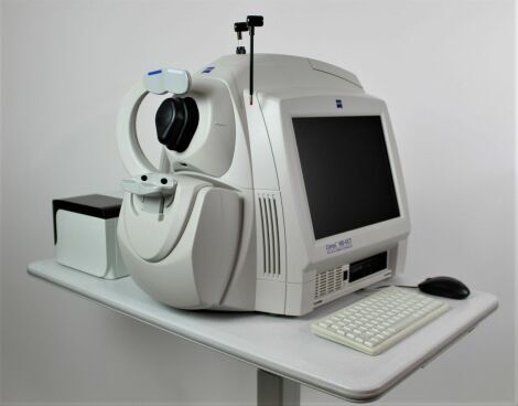
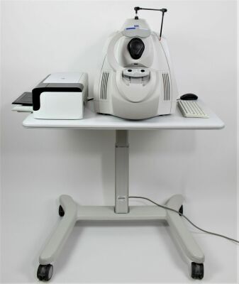
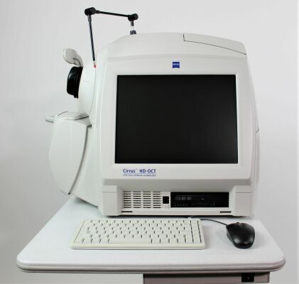
As the world’s leading OCT innovator, Zeiss has been at the forefront of industry-defining advancements that have made OCT standard of care. The Zeiss Cirrus HD-OCT 4000 enable examination of the posterior and anterior of the eye at an extremely fine spatial scale, without surgical biopsy or even any contact with the eye. The Cirrus builds on and refines the retinal imaging technology first introduced with the Zeiss Stratus OCT. Employing the advanced imaging technology of spectral domain optical coherence tomography, Cirrus acquires OCT data about 70 times faster (27,000 vs. 400 A-scans per second) and with better resolution (5 um x ~10 um axial resolution in tissue), compared to first-generation OCT technology. Cirrus acquires whole cubes of OCT image data, composed of hundreds of line scans, in about the same time as Stratus acquires a six-line scan. You can view these data cubes in three planes, or through three dimensions, giving you access to an extensive amount of retinal image data in one scan.
Indications for use
The Cirrus HD-OCT is a non-contact, high resolution tomographic and biomicroscopic imaging device. It is indicated for in-vivo viewing, axial cross-sectional, and three-dimensional imaging and measurement of anterior and posterior ocular structures, including cornea, retina, retinal nerve fiber layer, ganglion cell plus inner plexiform layer, macula, and optic nerve head. The Cirrus normative databases are quantitative tools for the comparison of retinal nerve fiber layer thickness, macular thickness, ganglion cell plus inner plexiform layer thickness, and optic nerve head measurements to a database of normal subjects. The Cirrus HD-OCT is intended for use as a diagnostic device to aid in the detection and management of ocular diseases including, but not limited to, macular holes, cystoid macular edema, diabetic retinopathy, age-related macular degeneration, and glaucoma.
Features:
-Quad core computer
-Eye tracking latest revision software
-Spectral domain imaging
-Cirrus cube analysis
-Macular normative data
-3D rending
-Acquisition auto focus
-Anterior segment analysis
-Optional FastTrac and up to 10 review licenses
Specifications:
HD OCT imaging:
-Methodology: Spectral Domain OCT
-Optical Source: superluminescent diode (SLD), 840 nm
-Optical Power: < 725 μW at the cornea
-Scan Speed: 27,000 A-scans per second
-A-Scan depth: 2.0 mm (in tissue), 1024 points
-Axial resolution: 5 μm (in tissue)
-Transverse resolution: 15 μm (in tissue)
Fundus Imaging:
-Methodology: Line scanning ophthalmoscope
-Live Fundus Image: During alignment and during OCT scan
-Optical Source: Superluminescent diode (SLD), 750 nm
-Optical Power: < 1.5 mW at the cornea
-Field of View: 36 degrees W x 30 degrees H
-Frame rate: >20 Hz
-Transverse resolution: 25 μm (in tissue)
Iris Imaging:
-Methodology: CCD Camera
-Resolution: 1280 x 1024
-Live iris image: During alignment
Computer:
• High performance multi-core processor
• Internal storage: > 80,000 scans
• CD-RW, DVD-ROM drive
• Integrated 15" color flat panel display
This Zeiss Cirrus HD-OCT 4000 is in excellent condition, meets manufacturers specifications and comes equipped with:
-6 Month warranty
-Power table and printer
-Manual and reference sheets
Return Policy
Items are sold as-is with no returns or refunds available unless explicitly stated.


 Get a CPS 2-Year Protection Plan starting at $5.99. Sold separately. Learn more